
Managing Necrosis from Hyaluronic Acid Filler Injection
Delays in treatment can result in severe scarring.
Key Takeaways
- Early recognition and treatment of vascular occlusion are critical after hyaluronic acid filler injection, as delays in managing skin necrosis can result in severe, persistent scarring.
- Hyaluronidase is the cornerstone of initial treatment for suspected HA filler–induced vascular occlusion and should be administered promptly, with additional treatment considered when clinical improvement is inadequate.
- Necrosis may initially resemble herpes infection, making vigilance essential when patients develop pain, swelling, discoloration, eschar, or other concerning skin changes following filler injection.
- Prevention and preparedness are essential, particularly when injecting high-G′ fillers into confined, high-risk areas such as the forehead and glabella.
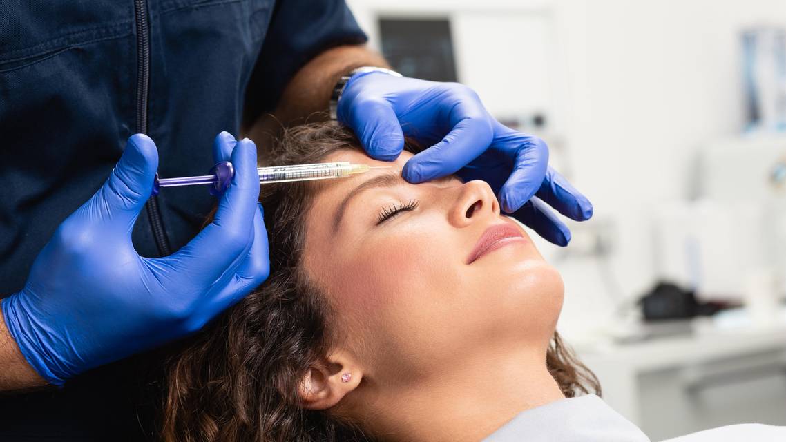
Hyaluronic acid (HA) dermal filler injections have become a popular aesthetic treatment in large part because of minimal immunogenicity and relative ease of use.1 The treatment is generally believed to be safe, but serious complications such as infection, skin necrosis due to vascular occlusion, and blindness occur on rare occasions.2,3 A 2025 retrospective study of more than 200,000 patients found 12 cases of serious complications—10 of which involved vascular obstruction.3
As rare as those cases are, it is imperative for the clinician to recognize them as quickly as possible and treat appropriately to minimize long-term skin damage. Hyaluronidase is the baseline treatment for skin necrosis due to vascular occlusion after HA dermal fillers.4,5 A 2015 consensus paper recommended immediate treatment with at least 200 units of hyaluronidase, plus more if improvement is not seen within 60 minutes.4 Adjunctive treatments can include nitroglycerin paste, an oral aspirin regimen, and other options, and once again, timing is critical.4
During those initial moments, differentiating between herpes and necrosis can be challenging. Any clinician injecting HA dermal filler should be vigilant for necrosis in these situations. The following case report illustrates a situation when hyaluronidase was administered immediately but further treatment for necrosis was delayed by an initial belief that the patient’s symptoms indicated herpes. The patient required nearly 2 years of subsequent treatments to resolve the resulting scarring.
CASE REPORT
A 43-year-old female patient sought to eradicate lines and wrinkles from her forehead and visited a medical spa for injections. A physician administered both neuromodulator and HA dermal filler injections in her glabella and forehead, and almost immediately, the patient experienced esotropia of her right eye, swelling, pain, and blurred/double vision. The doctor, upon recognizing the exotropia, administered 300 units of recombinant human hyaluronidase (Hylenex; Halozyme Therapeutics).
Suspecting retrobulbar involvement, the doctor sent the patient to an ophthalmology hospital, but by the time she arrived, her vision had normalized, and the ophthalmologist did not deem it necessary to administer retrobulbar hyaluronidase.
The patient then returned to the injecting doctor and received another 150 units of hyaluronidase—for a total of 450 that day—along with an intralesional steroid and was sent home.
The next day, the patient noted darkness in her skin and swelling in her eyelids. She returned to the doctor, who provided nitroglycerin patches, but those caused her to experience headaches.
Over the course of the next 2 days, the patient developed a very black eschar, and the lesion became exophytic. She developed further problems and breakdown of the skin, but the doctor advised that it was likely a complication with herpes. She then visited an emergency room, where a culture was performed to rule out herpes simplex virus; the culture was negative. A plastic surgery resident and a hyperbaric oxygen consultant examined her and discharged her, advising that the skin would likely heal on its own with the use of petroleum jelly.
When the patient’s condition had not improved after 2 more days, she visited our dermatology office, where she was diagnosed with necrosis of the skin (Figure 1) and treated in a hyperbaric oxygen chamber. She did not require any further hyaluronidase, and 9 days later, the skin had developed scabs (Figure 2).


Approximately 1 month later, the necrosis had resolved, but severe exophytic scars had developed, especially on the bridge of her nose (Figure 3 and Figure 4). Intralesional steroids were ruled out as an option due to the high concern of necrosis from particulate matter in this area, especially given the patient’s recent history. Topical steroids were discussed, but we decided to first try waterproof hydrocolloid wound dressings (DuoDERM; ConvaTec) in hopes that they may flatten the lesions. Finally, the patient was advised to consult with a plastic surgeon about three further options: shave removal, hydrosurgery, or CO2 laser ablation. In the meantime, she was advised to continue with hyperbaric chamber therapy.


Approximately 4 months later, the patient returned (Figure 5) and requested neuromodulator injections for her glabella. She was injected with 10 units of onabotulinumtoxinA (BOTOX Cosmetic; Allergan Aesthetics).

The patient returned to the office approximately 7 months after that with marked improvement in redness but was now concerned with the white scars and unevenness. She expressed interest in microneedling with radiofrequency and CO2 laser treatment.
We treated her glabella, forehead, dorsal nose, and nasal sidewall scars with microneedling with radiofrequency (VirtueRF, Cartessa Aesthetics), level 3, for 600 ms. We then used a CO2 laser (CoolPeel, Cartessa Aesthetics) with the following parameters:
- Power: 3 w
- Dwell time: 0
- Spacing: 600 µm
- Pattern: Square
- Size: 10 and 6 and 4
When the patient returned 8 weeks later, she felt that some of the scars had flattened, but white patches and plaques remained on her forehead (Figure 6). She underwent another round of microneedling with radiofrequency, this time at level 4, along with CO2 laser treatment with the following parameters:
- Power: 4 w
- Dwell time: 0
- Spacing: 450 µm
- Pattern: Square
- Size: 6

Immediately following those treatments, she also was injected with 12.5 units of onabotulinumtoxinA in the glabella and 7.5 units in the forehead.
The patient returned for the same treatments twice more, 8 weeks apart (Figure 7 and Figure 8).


Figure 8.
The final round of treatment included separate rounds of microneedling with radiofrequency at level 5: on the glabella/forehead/bridge of nose, and on the remainder of the face other than the scars. We also used the CO2 laser at 5 w on each of those areas.
The treatments ultimately achieved great improvement, and the patient was very satisfied (Figure 9).

Figure 9.
DISCUSSION
This case reinforced the importance of recognizing necrosis early and treating with hyaluronidase, nitroglycerin paste, and hyperbaric oxygen chambers. We developed a protocol because of this case in the event that this sort of complication ever occurs with any of our own patients. Sadly, it is not all that uncommon, but clinicians often do not recognize it immediately; I have seen similar cases also initially thought to be herpes.
The other lesson from this case was that clinicians must be very careful injecting high-G’ fillers into small spaces, especially on the forehead, because necrosis is a real entity.Whether you are a novice injector or a seasoned injector, these things can happen.
We must explain to every patient that this is an unlikely but possible side effect, and when performing these injections, we need to be ready and willing to treat necrosis immediately. The consequences of delayed treatment can be mitigated, but it can take years.
Ready to Claim Your Credits?
You have attempts to pass this post-test. Take your time and review carefully before submitting.
Good luck!
Recommended
 Injectables
Rethinking Delayed Inflammatory Reactions to Hyaluronic Acid Fillers
Gretchen W. Frieling, MDGretchen W. Frieling, MD