
Picosecond Laser Study Links Immediate Whitening to Skin Tissue Response
Key Takeaways
- Picosecond laser–induced immediate whitening (IW) corresponded with the spatial scale of acute tissue responses in normal human skin.
- Melanosome disruption occurred even without visible IW; slight IW was associated with vacuoles that could exceed basal cell size.
- Compared with a 755-nm nanosecond laser, picosecond irradiation produced more spatially confined vacuolization at matched IW endpoints but larger vacuoles when fluence at the dermoepidermal junction was matched.
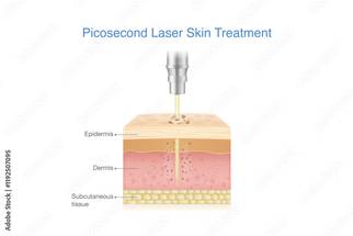
A new in vivo study offers a closer look at what immediate whitening (IW) may represent at the tissue level following picosecond laser irradiation.
Researchers publishing in Lasers in Surgery and Medicine, the study evaluated the relationship among IW, vacuole formation, and melanosome disruption after 755-nm picosecond laser irradiation of normal human skin.
Immediate Whitening May Reflect the Scale of Picosecond Laser Effects
The investigators applied a 755-nm picosecond laser under varying irradiation conditions and clinically graded IW responses. Histologic analysis quantified vacuole number and area, while electron microscopy evaluated melanosome morphology. The researchers also used a theoretical melanosome disruption threshold fluence model to estimate fluence at the DEJ and compared picosecond and 755-nm nanosecond laser irradiation under endpoint-matched and DEJ fluence-matched conditions.
Vacuole area increased with the degree of IW. With slight IW, some vacuoles exceeded the cellular scale, defined by basal cell area. In contrast, when no IW was observed, vacuoles remained below the cellular scale despite evidence of melanosome disruption. The findings characterized a shift from subcellular immediate responses without IW to cellular-scale effects with slight IW.
The theoretical analysis indicated that most tested irradiation settings exceeded the threshold for melanosome disruption. Compared with nanosecond irradiation, picosecond irradiation produced more spatially confined vacuole formation at matched IW levels but larger vacuoles when DEJ fluence was matched.
The study examined immediate responses in normal human skin, and the abstract does not report clinical outcomes in patients with pigmented lesions.
“IW reflects the spatial scale of picosecond laser-induced immediate responses and serves as a mechanistic indicator for disease-specific clinical endpoint selection,” the authors wrote. “These findings provide a framework for optimizing and standardizing picosecond laser treatment parameters.”
Source
Omatsu J, et al. Laser Surg Med. 2026. Doi:10.1002/lsm.70196